
The flexor tendons are surrounded by a sheath, extending from the base of P3 to the head of the corresponding metacarpal. This sheath is lined internally with synovial tissue, circumscribing a virtual cavity filled with synovial fluid.
Infection of this sheath is called a phlegmon.
Two modes of contamination can be distinguished:
- Direct inoculation by a vulnating agent: in the case of a septic bite or sheath wound, germs can spread rapidly along the tendon.
- Indirect inoculation by diffusion on contact with the sheath from a neighboring infection.
This is a serious, rapidly evolving infection that can leave serious after-effects if not diagnosed and treated early.
CLINICAL SIGNS
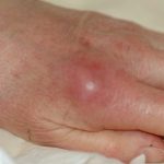
A few hours to a few days after the trauma (knife or screwdriver wound, prick, scratch, bite, etc.), swelling of the finger, pain along the finger path, in the palm or even in the wrist crease appear.
Extending the finger is unpleasant, and pressure on the cul-de-sac of the sheath is painful.
Later on, the finger becomes red and swollen, the pain is insomniac and extension of the finger is impossible, taking on a hook-like attitude. Nodes may appear, along with lymphangitis and fever. There are 3 successive stages:
stage1: exudative synovitis, which distends the flexor sheath; the fluid is dull or clear, abnormally abundant and the synovium is inflammatory.
Stage 2: purulent synovitis; damage to the synovium is irreversible, but the tendon is still intact.
Stage 3: infectious tendon necrosis.
THE TREATMENT
Treatment is always surgical, as antibiotics alone are unable to penetrate the tendon sheath.
Treatment involves excising the entire wound path en bloc.
- In stage 1, a proximal approach to the cul de sac enables sampling and washing of the tendon sheath. If there is pus in the sheath, a complete opening of the finger is warranted to allow removal of all infected or necrotic areas.
- In stage 2, the finger is opened along its entire length and a complete synovectomy is performed, respecting the mechanical continuity of the pulleys. Multiple washings are performed. Closure is incomplete to gain access to the infected site.
- In stage 3, partial or complete resection of the tendon is sometimes necessary, with subsequent directed healing or local flaps.
After surgical treatment, additional antibiotic therapy may be prescribed, depending on the severity of the lesion and the germ found.

SPECIFIC RISKS
Progress is favourable if treatment is carried out early.
If the infection is already significant, the secondary appearance of adhesions is responsible for stiffening of the finger.
In forms seen and treated too late, the tendon is necrotic and must be removed. The finger remains permanently hooked, and infection can lead to amputation.
