Preiser’s disease is defined by avascular necrosis of the scaphoid, occurring in the absence of trauma, fracture or pseudarthrosis.
The scaphoid is one of the largest on the carpus, located on the lateral side of the wrist, and is extremely mobile in all wrist movements, with fragile vascularization.
This pathology is extremely rare, and generally affects young adults.
Patients complain of vague pain on the outside of the wrist (on the thumb side). Clinical examination is rather poor, with occasional tenderness over the scaphoid and constant loss of strength.
Radiological images must be interpreted according to context:
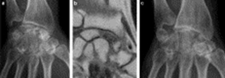
- Standard X-rays of the wrist, from the front and the side, with X-rays of the healthy side to compare the two images.
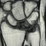
- An MRI scan will clarify the extent of the lesions,
- An arthroscanner will determine the state of the bone structure and, above all, the state of the cartilage of the scaphoid and radius: these two elements are important in deciding on the surgical option.
Stage I: normal radiological image; this is an early stage. MRI and bone scintigraphy are abnormal.
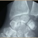
Stage II: contrast alterations in the scaphoid, predominantly at the proximal pole, close to the radius: the shape of the scaphoid is preserved.
Stage III: loss of scaphoid shape, sometimes fragmenting; the upper portion of the scaphoid is collapsed with an eggshell pattern, indicative of fracture.
Stage IV: severe collapse of the scaphoid, with images of osteoarthritis of the wrist between the scaphoid and the radius.
Traitement:
When the shape of the scaphoid is more or less preserved and its cartilage is still correct, bone grafting is performed: conventional or vascularized bone grafting.
When the shape and cartilage of the scaphoid are affected, resection of the first row of the carpus is the best option.
Treatment varies according to the stage of the disease, and your surgeon will give you the information you need.
