In general, carpal dislocations occur more rarely than fractures. However, it is possible for a dislocation to be accompanied by a fracture. The carpus is made up of ten or so bones, which may be dislocated or dislocated. Following carpal dislocation, wrist movement becomes impossible. Untreated injury can lead to relatively serious complications.
CAUSES AND COMPLICATIONS
Dislocations most often occur as a result of violent trauma. This could be a traffic accident (motorcycle), a fall from a great height, etc. The people most affected by carpal dislocations are young adults (20 and over).
Because of their bone strength, carpal bones are difficult to fracture, but can nevertheless dislocate. In the elderly, fractures are sometimes accompanied by dislocation.
In most cases of dislocation, it is the lunate that becomes dislocated. This can lead to nerve complications. The lunate can displace and compress the median nerve, whose branches run through the joint.
DIFFERENT TYPES OF CARPAL DISLOCATION
There are several types of carpal dislocation. These dislocations may be simple, i.e. without fracture of the carpal bones. In most cases, the lunate is displaced and lies alone in front of the other carpal bones. This is known as retro-lunar dislocation.
This type of dislocation may leave after-effects. Dislocations can also be accompanied by fractures. This may be a trans-scapho-retrolunate fracture-luxation (scaphoid fracture), or a fenton syndrome (capitatum fracture).
CLINICAL SIGNS
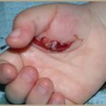
After a carpal dislocation, the wrist becomes deformed as a result of the swelling caused by the dislocation. If a hematoma and edema form, this may be a dislocation with complications. On palpation, the wrist is extremely painful. The patient may also feel a tingling sensation and have the impression that the hand is paralyzed.
ADDITIONAL TESTS
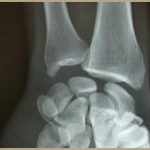
The doctor usually prescribes additional tests. These are mainly X-rays. Two x-rays are usually taken, showing the wrist from the front and the side. If the X-ray is not precise, or if the lesion is complex, the doctor may opt for a CT scan. This enables all the bones to be observed and their position precisely determined.
THE TREATMENT
In the event of an emergency, medical staff use external maneuvers to reduce the dislocation. If the dislocation is severe, the doctor will perform a minimally invasive procedure to repair the various damaged ligaments.
In the case of fractures, the surgeon will seek to replace the bones or bone fragments using osteosynthesis. In the case of trans-scapholunate dislocation, screws or pins are used to stabilize the bone tissue. During the operation, the median nerve may also be neurolyzed.
