
The scaphoid is the largest and outermost bone in the first row of the carpus (wrist).
Located on the thumb side, it is highly mobile and vulnerable to wrist trauma, and is the most frequently fractured bone.
Due to its particular location and vascularization, it tends to consolidate with difficulty.
After 6 months, a fracture that has not healed is called “pseudarthrosis”.
Pseudarthrosis means that the physiological mechanism of bone consolidation is “blocked”.
Scaphoid fractures generally occur in young (20-30 years), athletic men.
It occurs after a fall onto the extended wrist.
These fractures can develop into pseudarthroses for a number of reasons:
- poor initial immobilization
- Early interruption of immobilization
- Late diagnosis of scaphoid fracture
- vascular characteristics of the scaphoid
- Scherenberg fracture classification

SIGNS AND SYMPTOMS:
They are often very slight, which is the main problem, as the patient readily considers it unnecessary to seek medical attention for what appears to be a simple contusion. As a result, the injury is often overlooked at the time of the initial trauma.
Palpation of the area around the base of the thumb and compression in its axis are painful, as are forced movements of the wrist.
The diagnosis of pseudarthrosis may be made by chance on an X-ray requested during a 2nd trauma, occurring long after the one responsible for the scaphoid fracture, the initial accident having sometimes been forgotten by the patient.
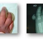
RADIOLOGICAL ASSESSMENT:
X-rays of the wrist in front and in profile, and specific scaphoid incidences (Schneck’s incidence).
On X-rays, it is sometimes difficult for a non-specialist to distinguish between a recent fracture and an old, non-union fracture (pseudarthrosis).
MRI or CT scans can confirm the diagnosis of pseudarthrosis and detect bone necrosis at the proximal pole of the scaphoid.
EVOLUTION:
Disorganization of the carpal bones, leading to early wear and tear (arthrosis) of the joints connecting them. Within 5 to 10 years, this osteoarthritis will lead to significant or even total functional incapacity of the wrist: stiffness, pain and loss of strength. It is therefore essential to diagnose and treat this pseudarthrosis before the onset of osteoarthritis.
Traitement:
Depending on the age and stage of the pseudarthrosis, your surgeon will suggest the appropriate treatment.
Alnot’s classification suggests 4 different stages according to which treatment will be proposed
- Stage I: linear pseudarthrosis with no change in the shape of the scaphoid, and no intra-carpal instability or misalignment.
- stage II: divided into IIA when the pseudarthrosis is stable, with the onset of bone resorption at the fracture line, and stage IIB when the pseudarthrosis is unstable, with loss of anterior substance and flexion of the proximal pole on the distal tubercle, leading to intracarpal misalignment with DISI (Dorsal Intercalated Segment Instability).
Stage III: corresponds to an unstable displaced pseudarthrosis with intra-carpal misalignment as in Stage IIB. This stage is divided into two sub-stages: Stage IIIA with stylo-scaphoid arthrosis and Stage IIIB with radio- and inter-carpal arthrosis.
The first three stages of this classification represent a single lesion left to its natural evolution.
stage IV: a special stage corresponding to necrosis of the proximal fragment, with stage IVA, where there is misalignment, and stage IVB, where there is radioscaphoid and intercarpal osteoarthritis.
If osteoarthritis is absent: we can treat the pseudarthrosis and attempt to consolidate the fracture by using osteosynthesis material (screws or pins) in the scaphoid to restore its anatomical shape and height (stage I-IIA).


If there is bone loss at the site of pseudarthrosis (stage IIB-IIIA), a bone graft from the pelvis or radius will be used, with or without osteosynthesis.
The same treatment is performed in stage IIIA, with the addition of a radial styloidectomy.
The scar is usually located on the anterior aspect of the wrist.
Bone harvesting, particularly from the pelvis, may cause pain for a few days, but does not leave any after-effects.
In some cases, a vascularized graft may be used, i.e. a bone fragment accompanied by its feeder artery, if the vitality of the scaphoid is impaired.
Depending on the technique used, the wrist will be immobilized after the operation for a period ranging from a few weeks to 3 months.
After this operation, which generally requires a 24-hour hospital stay, the rate of consolidation is high, and it is possible to recover near-normal wrist function in terms of strength and mobility.
In some cases, however, consolidation is not achieved, and further surgery is required.
If radiocarpal or intracarpal osteoarthritis is present (stage III and IV A): this is a “salvage” operation designed to restore a “certain” degree of mobility, halt the arthrosic process and eliminate pain. Depending on the stage of the SNAC, either :
- Resection of the first row of carpal bones. This is only possible in stage II. The scaphoid, lunate and triquterum are removed, and the intact capitate is articulated with the distal end of the radius.
- Partial arthrodesis of the carpal bones associated with scaphoidectomy. The diseased scaphoid is removed and, to leave the other bones “coherent”, they are fixed together with the aim of fusing them using a specific material. Screw fixation, staple or osteosynthesis plate (locked xPode plate).
Stage IVB pseudarthrosis: wrist arthrodesis, wrist prosthesis, neurectomy
Rehabilitation begins after the immobilization period. Rehabilitation should restore joint range of motion, with secondary strengthening of clamping strength. Sports activities can only be resumed after the 6th month.
