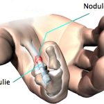
Dupuytren‘s disease is linked to thickening of the palmar aponeurosis (“palmar aponeurosis”), a structure located under the skin of the palm and fingers. It is sometimes accompanied by digital, palmar or mixed nodules and retraction of one or more fingers, limiting their extension. The evolution of this retraction depends on the type of involvement and the aggressiveness of the disease.
Clinically, we find digital, palmar or digitopalmar flanges. Hard nodules and pad-like depressions are always found in the palm of the hand. There are also forms with pads on the dorsal surface opposite the proximal interphalangeal joints.
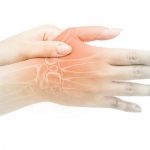
The 4th and 5th fingers are the first to be affected in the majority of cases, although all fingers may be affected, with bilateral lesions in half of cases. Exclusive involvement of the 5th finger is said to be difficult and recurrent.
Diagnosis of this disease is clinical and does not require further investigation.
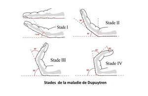
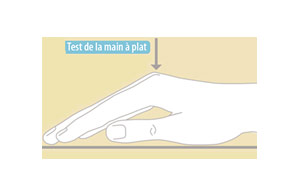
The evolution of the disease is monitored using the Tubiana classification, and the indication for surgery arises when the hand-table test is positive: “the hand can no longer lie flat on the table”.
THE TREATMENT:
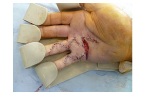
The aponeurectomy consists in removing the pathological tissue. This is a delicate operation, as the vascular and neural pedicles are in contact with the flanges to be removed.
If the retraction is significant and long-standing, the joints become stiff and it is not always possible to regain full extension. In the case of significant palmar retraction, the palm of the hand must be left open. Substance loss is treated by directed wound healing, with regular dressings for 2 to 3 weeks. Skin grafting may be necessary when the skin is invaded, or in the case of recurrence.

Your surgeon will advise you on the best course of treatment, depending on the stage of your Dupuytren’s disease, to restore as much digital extension as possible, while avoiding recurrence as far as possible.
It may be necessary to wear a dynamic extension orthosis for the affected finger for two months after the operation, at night, to avoid flexion scarring.
Rehabilitation is started as soon as the skin has healed
